
Distal Radius and Ulna Fractures
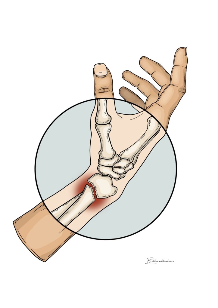
The ulna and radius are the two bones that make up the forearm, and in their distal portion, they form the wrist joint along with the carpal bones.
Distal radius fractures are the most common forearm fractures and usually result from a fall onto the hand. Due to their importance in the wrist joint, accurate diagnosis and treatment are essential to avoid long-term complications.
The usual injury mechanism for distal radius fractures is a fall onto the hand, although they can occur in other traumas such as sports accidents, traffic accidents, etc. The ulna is often fractured in the context of radius fractures during a fall, which can cause instability of the distal radioulnar joint. This instability must be treated at the same time as the radius fracture.
Wrist pain
This usually increases with wrist flexion and extension movements or when direct pressure is applied to the fracture.
Swelling
The affected area is usually swollen as a result of the fracture.
Visible deformity
Due to the superficial location of the radius and the deformity that is common in fractures, visible deformity of the distal forearm is frequent.
Bruising
Due to the bleeding that occurs after the fracture, bruising is common around the wrist.
Decreased range of motion
Diagnosis of Distal Radius and Ulna Fractures
Diagnosis is usually made through medical history and physical examination, supported by wrist X-rays where the fracture line is typically visible.
Occasionally, a CT scan is necessary to visualize the fracture lines and bone fragments, aiding in treatment planning and decision-making. For some types of fractures, wrist arthroscopy is often recommended to diagnose and treat associated injuries.
Treatment of Distal Radius and Ulna Fractures
Treatment will depend on the patient’s characteristics and the fracture itself, but generally includes the following options:
Splint Immobilization
In cases of minimally displaced fractures with favorable characteristics, immobilization with a forearm cast and radiographic monitoring can be performed.
Open Reduction and Internal Fixation
In cases of displaced or intra-articular fractures, surgical treatment with open reduction and internal fixation is usually recommended.
Arthroscopic Reduction and Internal Fixation
In cases of intra-articular fractures with multiple fragments, arthroscopic reduction of the fragments is recommended to restore the articular surface and reduce the risk of future complications. It also allows for the diagnosis and simultaneous treatment of associated injuries, such as those of the triangular fibrocartilage complex (TFCC) or the scapholunate ligament.
Would you like a valuation? Make an appointment with us.
Every case is unique. If you need an expert opinion on your situation, we will be happy to help you. Fill in the form and we will contact you to give you a personalised assessment and, if you wish, arrange an initial appointment.